History of Neuroimaging
Presented to the ASN from the Archives by William McKinney. Edited and adapted for the ASN website by Rohit Bakshi.
Presented at the ASN 20th Annual Meeting, Puerto Rico, 1997, by Anne M. Watterson, Michelle G. Flye, The Dorothy Carpenter Medical Archives, and William McKinney. Portions also taken from Jack Greenberg’s history of neurology and neuroimaging – written for the 50th Anniversary of the American Academy of Neurology 1895-1973: The beginning of modern neuroimaging.
1895-1973: THE BEGINNING OF MODERN NEUROIMAGING
In 1895 Wilhelm Roentgen, a physicist, demonstrated the first radiograph and opened a new window to medical diagnosis. His revolutionary discovery set the stage for even more amazing advances in the imaging of human disease. William Oldendorf, MD traced the development of neuroimaging in his Wartenberg lecture to the Academy of Neurology (AAN) in 1978. He emphasized the diversity among the scientists and medical specialists who contributed. Walter Dandy, a neurosurgeon, first performed ventriculography and pneumoencephalagraphy (PEG) in 1918 and 1919. Moniz, a neurologist, accomplished the first cerebral arteriogram in 1927. Oldendorf himself developed the basis for computerized tomography (CT) in 1961 and the technique was applied to clinical diagnosis by an electrical engineer, Hounsfield, in 1973. Finally, magnetic resonance imaging (MRI) was introduced. In the mid 1970s, Lauterbur, a physicist, published the first spatially differentiated MR images and Damadian, an internist, published on MRI tumor detection.
1953-1975: THE ROLE OF WILLIAM OLDENDORF - NEUROLOGIST AND NEUROIMAGING PIONEER
"My most memorable experience involving the ASN was receiving the Oldendorf Award from Bill Oldendorf himself. The meeting was held in San Juan, and Bill and I flew there together from Los Angeles. Despite the fact that we were both members of the UCLA Neurology Department, we had hardly met before the trip because I was a resident and he was a senior faculty member running his research lab at the Wadsworth VA Hospital. After that meeting, we became close friends and I always trusted his advice, both in areas of imaging research, as well as in important professional decisions. He was a man for all seasons in the world of imaging and in neurology. I miss his good humor and quick wit and fondly remember the part the ASN played in initiating our friendship” -John C. Mazziotta, M.D., Ph.D., Past President, American Society of Neuroimaging.
Oldendorf had trained with A.B. Baker in the early 1950's in one of the few programs requiring neurology residents to perform arteriograms and PEG’s on their own patients. From 1953 to 1955 Oldendorf did all these invasive studies on his patients at the University of Minnesota Hospital. Unsatisfied that these traumatic, tedious tests provided only limited and indirect information about the brain, he strived for something better. In 1961 he published a description of a new instrument, modeled in his basement and elegantly simple, the first to incorporate the principles and hardware eventually used by modern CT scanners. Although Oldendorf’s idea was patented in 1963, manufacturers of standard x-ray equipment dismissed the new technique as impractical. A letter from one company ended: “Even if it could be made to work as you suggest, we cannot imagine a significant market for such an expensive apparatus which would do nothing but make a radiographic cross-section of a head.”
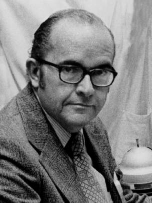
William H. Oldendorf, M.D., 1925-1992
Dr. Bill Oldendorf, a founding member of the ASN, was widely recognized as one of the original founders of the principles of computed tomography. Oldendorf’s studies in the late 1950s and early 1960s were acknowledged by Sir Godfrey Hounsfield in his own work that led to the invention of the X-ray CT scanner and the Nobel Prize for medicine in 1979.
Oldendorf’s work was invaluable to researchers in the field of neuroimaging, despite his inability to interest X-ray equipment manufacturers in his invention. In 1975, Oldendorf and Hounsfield were awarded the Albert and Mary Lasker Award for the conception of the principles that led to the development of computed tomography. Oldendorf’s work has also been applied to the areas of PET and SPECT imaging.
-Adapted from: Mazziotta JC, Collins RC. William H. Oldendorf, 1925-1992. Ann Neurol 1993; 33:331.
In recounting a history of his life in neuroimaging to the American Society of Neuroimaging (ASN) in 1992, Oldendorf discussed Godfrey Hounsfield and his successful introduction of the CT scanner. “He was kind enough to mention my 1961 paper as the only preexisting attempt to do the same thing that he did. Although the original CT picture was crude, it was obvious that it was a revolution.” Hounsfield was awarded the Nobel Prize for this work, along with Alan Cormack, a physicist.
In May 1973 Oldendorf brought Polaroid photographs to the United States made with the second clinically active CT in England and presented them at the Los Angeles Neurological Society. Again his efforts received little interest! Two years later he appeared on the same program (this time with much better slides) to provide an update on this technology to his colleagues. He overheard one neurologist ask another about the evening’s lecture. Upon hearing the topic would again be CT, the second fellow responded: “what, again?”
In 1975 James Toole, MD chairman of the Ad hoc committee on Neuroimaging of the American Academy of Neurology (AAN) wrote to Oldendorf regarding his opinion of the effect of CT scanning on the practice of neurology. Oldendorf responded:
“I think the EMI CT scanner in particular is going to threaten the clinical neurological world. I have recently seen the Mark 3 of the head-scanning unit that will be shown by EMI at the RSNA in November. It is far in advance of any of the head-scanning units now in use. Its display of brain structure and tissue density is so elegant that it actually obviates most of the detailed clinical analysis we have sweated over for so many years. In the old days when the general practitioner wanted an analysis of a possible brain problem, he would send the patient for an EEG and use the interpretation of the tracing as a neurological consultation. This was, of course, foolhardy. But when he now sends the patient for an EMI scan, it really is beginning to fulfill much of the function of a neurological consultation. As ultrasound appears, and I am certain that it will appear in full force in the next few years…to continue to hitch our wagon to the star of EEG is becoming more and more anachronistic each day. We must embrace these new techniques. If we don’t get on board, we will be left at the station.”
1975: CREATING THE POLITICAL WILL FOR THE ASN
In 1972 Buffalo Neurologist, Dr. William Kinkel, attended a refresher course on neuroradiology at Albert Einstein Medical Center in New York. Dr. James Bull, a well-known British neuroradiologist introduced Godfrey Hounsfield. They flashed an image on the screen, the first CT scan shown in the United States. So stimulated by this image, Kinkel went to the EMI company in England to look at the CT scanner. Hillier Baker, a radiologist at the Mayo Clinic, preceded him. In 1972 the Dent Neurologic Institute in Buffalo, New York, which Kinkel headed, then obtained the third scanner in the United States after Massachusetts General Hospital and the Mayo Clinic. In 1972 Dr. William Stuart of Atlanta, Georgia visited the Massachusetts General Hospital to see a new piece of equipment that “was going to revolutionize the practice of neurology.” He returned to Atlanta with Polaroid images from the “primitive CT scan of that period.” The Atlanta neurologists bought the 16th scanner off the EMI assembly line. In 1973 Dr. Jack Greenberg saw pictures of the first CT scan and related to a neurosurgical colleague that he “ . . . had seen Valhalla! The ventricles of the brain were visualized without injecting air!” The administrator of his hospital agreed to buy this technology, but the response of the radiology department, that “CT is a passing fancy,” frightened the administrator and he refused to buy the machine. The neurosurgeon boldly approached the hospital administration with a plan for a group of physicians (neurosurgery, neurology and others) to come up with the necessary $350,000. They introduced the first CT scan into Pennsylvania and the twenty-ninth in the country. In 1975, the MediMaine Health Associates, a neurologic group headed by Francis Kittredge MD, developed the first CT facility in Maine. (In 1985, they also brought the first MR facility to Maine and the first high-field MRI system to New England.)
At the Executive Board meeting of the American Academy of Neurology in Bal Harbor, Florida, April 30, 1975. Dr. Toole sent a letter to Dr. Floyd Davis, then chair of the special courses of the AAN requesting that CT and ultrasound courses be added to the program of the annual meeting. Toole (in another letter dated May 20, 1975 to Joseph Foley M.D., President of the ANA) writes, “As I talked with Bob Fishman (AAN President) I found myself appointed Chairman of an ad-hoc imaging subcommittee which is to report in December to the AAN.” In this letter Toole strongly requested that the ANA appoint a committee to represent the neurologist’s point of view regarding new imaging procedures. “A number of neurologists have involved themselves in imaging and a number more would, were they to be encouraged by their parent neurological association. The practice of neurology will be vastly changed by these new methods which neurologists should be intimately concerned with.”
Toole’s letter provoked a strong resolution by The American Neurological Association (ANA). The ANA celebrated its centennial meeting in New York City in June, l975. They hailed 100 years of progress in neurology. The Neuroimaging Commission on Neurology’s report concluded that the new methods for imaging the nervous system would greatly reduce the need for neurologists. “A CT scan can give a more accurate localization in far less time than a neurologist. Ultrasound of the carotid arteries can localize and indicate the degree of stenosis more accurately than a clinician with a stethoscope. The portent for the future is that neurologists who rely exclusively on their wits and their pins and hammers, unaware the machine age has finally come to neurology, may become obsolete. Those with an eye on the future must involve themselves in the supervision and interpretation of the new diagnostic procedures.” During the business session, ANA President-Elect Fred Plum recognized Bill Oldendorf’s seminal contribution that led to CT scanning. “It may be that some neurologists will adopt as their own, this pioneering work.” As the last act before adjourning the business meeting on June 4, l975, it was resolved:
“Because neurologists have traditionally been concerned with neurodiagnostic procedures and their interpretation, because the advent of new techniques for imaging the brain and spinal cord, for measuring its blood flow and neural function, are of greatest importance for care of patients with neurologic disorders and for teaching and research, therefore the Association wishes to emphasize that neurologists must be involved in the decision making, performance, and interpretation of these procedures. Neurologists must also be intimately involved with planning, implementation, and execution of programs designed to teach these new techniques to physicians, technicians, and students. This plants the flag of neurology deep in the territory of imaging procedures for all to see and it may become the rallying point of neurological clinicians as they seek to protect the interests and future of their specialty.”
Later in December 1975 the AAN Board accepted the resolution passed by the ANA in June 1975 and also accepted their Ad Hoc Committee on Imaging’s report that included the statement: “Neuroimaging is an integral part of the practice of neurology, that requires broad knowledge of neuroanatomy, neuropathology, pathophysiology, and clinical neurology. Proper interpretation of neuroimaging requires knowledge of the effects of disease on the neuroimaging examination, its indications, performance and interpretation.” The Ad Hoc Committee consisted of James Toole, William Kinkel, William Oldendorf, William Stuart, Maurice Van Allen, James Perry, John S. Meyer, William McKinney, A.B. Baker, Robert Fishman (ex officio), Peritz Scheinberg , Robert Joynt and Jack Greenberg.